Odisha State Board CHSE Odisha Class 11 Psychology Solutions Unit 2 Perceptual Process Long Answer Questions Part 2.
CHSE Odisha 11th Class Psychology Unit 2 Perceptual Process Long Answer Questions Part-2
Long Questions With Answers
Question 1.
What is Neuron? Describe the structures and functions of neurons, flow does it differ from a cell?
Answer :
Building Blocks of the Nervous System:
Neurons are basic units of the nervous system. These are the nerve cells that actually process information. The human brain contains about 100 billion neurons. The average neuron is as complex as a small computer and has as many as 10,000 physical connections with other cells. Most neurons are created very early in life, but their shape, size, and connections can change throughout the lifespan. On the whole, the way the neurons function reflect the major characteristics of the nervous system.
Basic Structure of a Neuron:
Not all neurons are alike. They are specialized to handle different information processing functions. However, all neurons have some common characteristics. In general, every neuron has the following structures
- Cell body or soma
- Dendrites
- Axon and
- Terminal buttons or axon terminals
(See fig. 4.1)
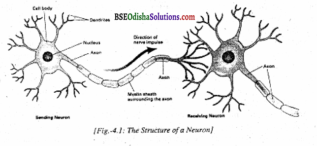
Cell body or Soma:
The cell body or ‘ Soma’ is the enlarged head of the neuron. It is enclosed by the cell membrane. The cell body contains the nucleus of the cell and cytoplasm which sustains its life. Some are the head side of the neuron. It uses oxygen and nutrients to generate energy. Its shape varies depending on the type of neuron. Generally, neurons transmit information in only one direction, that is, from the dendrites through soma to the axon to the terminal buttons.
Dendrites:
Dendrite is the branching fiber from the cell body. A neuron receives information at one end and sends out messages through the other. The part of the cell which receives incoming signals is called a dendrite. The dendrites receive nerve impulses from adjacent neurons or directly from sense receptors and conduct them to the cell body.
Axons relay or send impulses from the cell body to other neurons or to muscle tissue. The very word ‘dendrite’ came from the Greek word ‘dendron’ which means ‘tree’. So dendrites of a neuron look very much like trees. Dendrites are extended from the cell body. Dendrites increase the neuron’s surface area, allowing each neuron to receive input from many other neurons.
Axons:
Two types of extensions are found in the cell body. The short extensions from the cell body are called dendrites. But the longer single-branched extensions are called the axon. It is that part of the neuron which carries information away from the cell body to other cells. Each neuron has only one axon.
The point in the axon nearest to the cell body is called the axon hillock. Axons may have some branches which are called axon collaterals. Axons have two coverings. Of course, these two coverings are not found in every neuron. The outer boundary of the neuron is called the membrane. The membrane serves as a barrier for the neuron.
In some axons, there is a fatty white sheath called the myelin sheath. Axons having myelin sheath are called myelinated axons and which do not have it is called unmyelinated axons. Axons without myelin sheaths are not very good conductors of electricity. With the insulation of myelin sheaths, axons transmit electrical impulses and convey information much more rapidly.
Another covering is found in axons of neurons exclusively outside the brain and spinal cord. It is called a neurilemma. Neurilemma is a very thin covering that takes part in regeneration. If a neuron outside the brain and spinal cord is damaged, it can be regenerated. But the neurons of the brain and spinal cord can not be regenerated, as they do riot have neurilemma in their actions. Once these highly specialized cells are damaged, they are damaged forever.
Terminal Buttons:
An axon conducts information along its length which can be several feet in the spinal cord and less than a millimeter in the brain. At the far end of the axon, some swollen and bulb-like structures are there which are called terminal buttons. Through these buttons, stimulation passes to Astrocytes and oligodendroglia are two important glial cells.
Astrocytes produce chemicals that neurons need to fulfill their functions. On the other hand, astrocytes help control the chemical composition of the fluid surrounding neurons. The main function of oligodendroglia is to provide support to axons to produce myelin sheaths.
Functions neurons:
The main function of the neuron is to communicate messages of stimulation in the form of nerve impulses. Our behavior is only possible through the flow of nerve impulses. Near about 10 billion neurons fire in our brain. They send and receive various nerve impulses. This is the communicative function of the neuron.
Sensory or afferent neurons come from receptors and go to the brain and motor (efferent) neurons go to muscles or glands. The inter-neurons are the linking neurons. An electrochemical reaction occurs inside when a neuron is adequately stimulated. Neurons fire or do not file like a gun. There is no in-between stage. This is called the all-or-none principle.
All neurons follow this principle. They are either off or on. Now let us see how neurons serve their communicative functions and how nerve impulses or nerve energies are formed. The neuron contains intracellular fluid. The fluid on the outside of the neuron is called the extracellular fluid. In between these two types of fluids, there is a cell membrane.
The fluid contains many dissolved substances. Many chemical substances are broken into pieces when they dissolve in water or any fluid. Ions are electrically charged particles when dissolved. The electrical charges are negative or positive and are carried out by ions. As you know, positive and negative electrical charges attract each other.
But only positive electrical charges or negative electrical charges repel each other. Since ions are found in both extracellular fluid and intracellular fluid, the same thing happens in a neuron. (See fig. 4.3) A neuron works to maintain its resting potential. It does not come automatically.
When a neuron is in a resting state, there is a negative electrical charge of about – 70 million votes. Ameli volt one – thousand of a volt. This is called the resting potential of the neuron. The neuron can be best compared with a battery with the inside of the neuron representing the negative pole and the outside of the neuron representing the positive pole (Koester, 1991).
When a neuron is stimulated by externals like heat, light, or sound, the message arrives, and the positively charged ions outside the neuron rush inside the neuron at rates as high as 100 million ions per second. This sudden arrival of positive ions inside the neuron causes the charge to change from negative to positive.
When it reaches a critical level, an electrical nerve impulse known as action potential travels down the axon of the neuron. The very term ‘action potential’ is used to describe the brief wave of positive electrical charge which sweeps down the action.
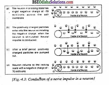
Generally, an action potential lasts only about 1/1000th of a second. When a neuron sends an action potential, it is commonly said to be ‘ firing’. The action potential abides by the all-or-none principle. Once the electrical impulse reaches a certain intensity, it fires and moves all the way down the axon without losing any of its intensity.
Again, the axon potential moves from one end of the axon to the other. After the nerve impulse has traveled, the positive ions are pumped out of the state. It becomes ready to fire again. The flow of the nerve impulse depends upon the diameter of a particular neuron. A nerve impulse is carried out speedily through a larger diameter and slowly through a smaller diameter.
Absolute Refractory Period:
After the action potential is transmitted by the neuron, it takes rests for a brief period of time. The neuron can not be fired again immediately, no matter how much stimulation it receives. It is just like reloading the gun after each shot. This time span just after carrying action potential during which the neuron is inactive is called the absolute refractory period.
During this period, the neuron is in resting potential. This time span of resting time is usually less than 1/1000th of a second. An action potential can not be produced during the absolute refractory period. When this span is over, again the neuron can cany a nerve impulse. The absolute refractory period is followed by a relative refractory period during which a strong stimulus can make the neuron active.
Threshold Point:
Weak stimuli can not produce an action potential in a neuron. Therefore, a stimulus of a certain strength is needed to produce an action potential. So the point at which a stimulus triggers an action potential is called the threshold of a neuron. Different neurons have different thresholds of excitation. Generally, the threshold point of each neuron is fairly constant.
Cell:
The nervous system of a living organism is made up of cells. A cell may be defined as a unit of living material. All can live independently by synthesizing within themselves substances from the nutrition absorbed from their environment. A cell contains living material called PROTOPLASM which is surrounded by a membrane called plasma membrane or the cell membrane.
![]()
Question 2.
Define AH or None law. Neural transmission or synapse?
Answer:
AH or None Law:
According to this principle, the nerve fibers respond completely or not at all. The stimulus has to be a minimum strength for the nerve to react. A weak stimulus will excite a few nerve fibers and we may not have sensory experience at all. But a strong stimulus will excite a larger number of nerve fibers resulting in a more intense experience. We can take the lighting of a match stick for illustration.
We must strike it with a certain amount of force to ignite the powder. If greater power is striking is exerted, the match flame will not be brighter. Beyond the minimum pressure necessary, any extra effort does not add to the brightness of the flame. This is exactly what happens with the stimulation of a nerve fiber. When such an explosion takes place, the nerve fiber is ready for another charge in a fraction of a second.
This principle is called the ‘all-or-none law”. Further, the nerve impulse is an electrochemical stimulation, which does not decrease in its intensity as it travels through the axon. If an axon carries any nerve impulse at all, the impulse continues to maintain the same strength throughout its travel in the axon until it reaches the terminal buttons.
The speed of a nerve impulse depends on the diameter of the axons. The larger the diameter, the greater is the speed. The strength of the nerve impulse depends upon the nature of the axons. It must be remembered that the dendrites and the cell body of a neuron do not obey the all-or-none principle. Only this principle is applicable to axons. So the axon is only governed by this law.
Neural Transmission:
No doubt, our mental functions stem from biological functions. In turn, they also influence our biological activities. Neural activity is biological activity. Neural activity is the biological medium in which all our psychological processes occur. Therefore, it is necessary to gain preliminary knowledge about how neural impulses travel from one part of the biological system to another. Not only the neural impulse travels within a neuron, but it also travels from one neuron to the other. The two major parts of the neural transmission are
- communication within a neuron (action potential) and
- communication between neurons (synaptic transmission).
We have already discussed how neural impulses travel from one neuron to another neurons.
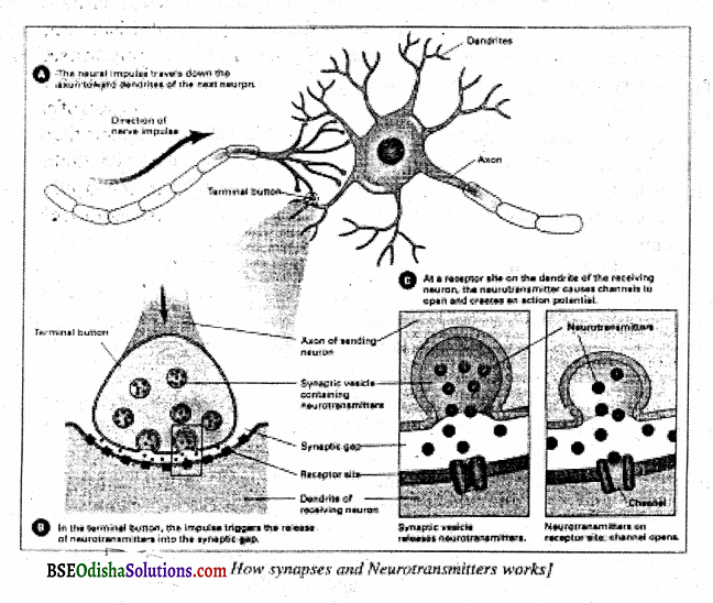
Synapse:
A synapse is a gap or junction which is generally found between the axon tip of one neuron and the dendrite of another. Neurons never touch each other. The nerve impulses are transmitted chemically across a small gap between the neurons. This type of contact provided between the axon of one neuron and the dendrites of another neuron is very interesting and significant.
The gap is very minute so that the conduction can easily go on. It is the synapse that makes our motor learning possible. When an impulse arrives at the end of an axon, electrical conduction in the axon is changed to chemical transmission. The tiny sacs in the terminal buttons of the axons, called synaptic vesicles release a transmitting substance called “neurotransmitters” which can the message to the other neuron.
Before the electrical impulse across the synaptic gap, it must be converted into a chemical signal As their name suggests, neurotransmitters transmit or carry information across the synaptic gap, it must be converted into a chemical signal. As their name suggests, neurotransmitters transmit or carry information across the synaptic gap to the next neuron.
When a nerve impulse reaches the end of the axon, it triggers the release of neurotransmitters which flood the synaptic gap. The dendrites of the receiving neuron come in direct contact with these neurotransmitters and receive the message. The receiving neuron will experience a change if the neurotransmitters are sufficiently stimulated, the received impulse will be conducted within the neuron and will be relayed from neuron to neuron until the message is completed.
It is estimated that the billions of neurons in the brain have trillions of synapses. There are many neurotransmitters. Each of them plays a specific role and functions in a specific pathway. Some neurotransmitters stimulate or excite, whereas, others inhibit neurons from firing (Bloom, Nelson & Lazerson, 2001). some neurotransmitters are both excitatory and inhibitory.
Most neurons secret only one type of neurotransmitter, but many different neurons are simultaneously secreting different neuro-transmitters into synaptic gaps of a single receiving neuron. Researchers have identified more than 50 neurotransmitters, each of which has a unique chemical makeup. Here, let us consider the functions of some neuro-transmitters which have major effects on our behavior.
Question 3.
Describe the structure and function of the spinal cord.
Answer:
Central Nervous System Structure and Function:
The central nervous system is well protected in the bony case of the skull and spinal column. It is divided into the spinal cord and the brain. The sensory or efferent neurons carry impulses from receptors into the central nervous system. The motor or efferent neurons cany impulses originating in the central system outward to effectors.
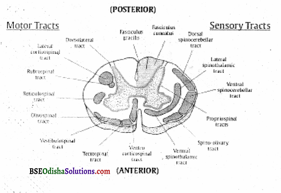
The Spinal Cord-Structure:
The spinal cord is a continuation of the brain below the medulla. It has a simple structure compared to the brain. It presents the same structure from angle to angle. A cross-section of the spinal cord at any level reveals the same uniform structure. The different parts of the spinal cord are connected to the brain.
The membranes cover the spinal cord and separate the fine neural tissues from the bony box of the central nervous system, lire outer, middle and inner membranes are called dura mater, arachnoid, and parameter respectively. The spinal cord has a rich acroterial blood supply. In a section of the spinal cord, we find grey matter in the central region and white matter in the peripheral region. The grey matter consists of millions of cell bodies of neurons.
The white matter consists of processes of neurons, that is, of axons and dendrites. In a six-foot man, the spinal cord is about the diameter of a little finger and 45 cm long. We do not find the spinal cord in most of the primitive forms of animal life. But when we go higher in the scale of evolution the nerve cells are found to be gradually combining into clusters and groups. These nerve clusters grow up the first step towards the evolution of the spinal cord and brain.
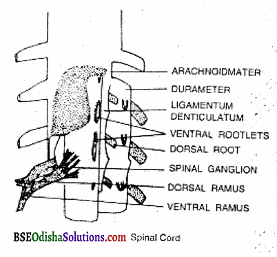
Connected to the spinal cord are thirty-one pairs of peripheral spinal nerves. In each nerve thousands of individual axons are bundled together. Some of these have sensory and some motor function. The sensory branches of the spinal cord enter the cord at the back of the dorsal portion.
The sensory branches cany into the spinal cord impulse generating in the sensory receptors in the skin, joints muscles, and viscera. After synaptic connections in the cord, the sensory activity runs toward the brain. On the other hand, the motor branches of the spinal nerves leave the front or ventral part of the cord. They control the axons of nearby muscles and glands.
Functions of Spinal Cord:
Complete transaction of the spinal cord proves that the spinal cord has communicative and integrative functions. Observations of patients show that if the cut of the spinal cord is above the level of exit of the spinal nerves to the arm and legs, the outcome is quadriplegia. Similarly, if the cut is below the arm level, but above the leg level, the person suffers from paraplegia.
In both cases, there is no recovery. There is complete anesthesia and permanent paralysis of the parts of the body below the level of the cut. Why there is paralysis? Because the sensory stimulations cannot reach the brain. The motor impulses also cannot come out because of this cut in the spinal cord.
So though voluntary movements in the parts of the body before the cut are absent, reflex actions are not lost. The presence of Kneejerk refer and the reflex arising out of the pinching in such patients indicate the integrative capacity of the spinal cord and also its capacity to respond adequately to simple stimuli and by acting differently to different stimuli.
The last act shows a crude kind of decision-making. However, the capacity to perform these actions upon command is lost in paraplegics. The moderating influences of the higher center are also lost. In normal people, the reflexes are under some sort of descending inhibition from the brain. But in these types of patients, the reflexes are relatively larger and more sudden.
![]()
Question 4.
What do you mean by reflex action and define its type or characteristic?
Answer:
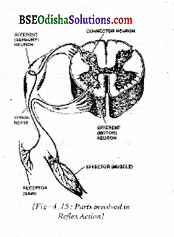
Reflex Action:
The reflex action is a very simple type of response that occurs automatically without our voluntary knowledge. According to Woodworth, “It is a direct muscular or glandular response to a sensory stimulus.” The examples of reflex actions are many. If someone puts his finger inside the mouth of a new baby, he starts sucking it. When a mosquito comes in front of someone’s eye, it is automatically closed as a protective measure.
Aeroflex action is very quick and rapid because it proceeds directly from the spinal cord. It does not go to the brain while in other activities the sensory stimulation is carried over to the brain through the spinal cord. The brain, in turn, sends out the message for making a particular response. That is why there is some delay in making these responses to activities other than reflex actions.
Types of Reflexes:
Basic reflexes, postural reflexes, segmental reflexes, inter-segmental reflexes, and spinal reflexes, come under different types of reflexes. There are also muscular reflexes such as knee jerks, the withdrawal of the hand, coughing, sneezing, swallowing, crying, and grasping reflexes. Glandular reflexes such as the flow of saliva, and the flow of tears are also notable.
Characteristics of Reflex Action:
The reflex actions are involuntary, unlearnt, and innate in nature like winking, knee erk, withdrawing leg from painful stimulation, sneezing, etc. The individual has no control over such reflexes. They occur very abruptly within a fraction of a second and also terminate very quickly. The involuntary or primary reflexes are purely universal.
The same type of avoidance of the pin pricking, grasping, winking and pupillary reflex is found in almost every person in the world. The reflexes are unlearnt behavior. Most of the behaviors of neonates are expressed in terms of reflexes. In babies, these reflexes are found in their original form.
As they grow up, the number of reflexes decrease and learned behavior increase. In other words, reflexes decrease with age. But there are certain reflexes that continue throughout life, like a knee-jerk, eye blink, etc. Finally, reflexes protect the individual from danger and help in maintaining the safety and welfare of the organism. Reflexes have, therefore, many positive values.
A reflex action is different from an ordinary action. In ordinary action, sensory stimulation in the form of electric waves is sent to the brain through the spinal cord. The brain then sends the message about the response to be made through the effectors. But in the case of reflex action, the message does not go to the brain. The spinal cord controls it. So the reflex action follows the spinal cord.
Reflex Arc:
The structure through which reflex action takes place is called the reflex arc. It involves the sensory nerve, spinal cord, and motor nerve. Take the example of withdrawing the foot when the pin is pricked. Sensory neurons cany impulses to the association.
Question 5.
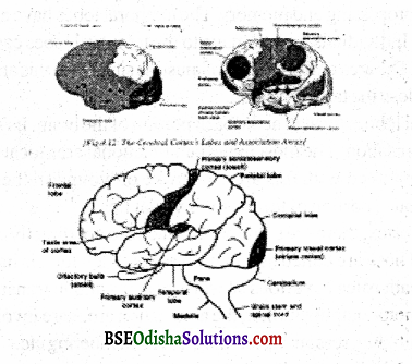
How many lobes are there in the brain? Describe their location and function.
Answer:
Four lobes, in the brain and these, are:
Frontal lobe:
It is located in front of the nearly vertical fissure of Rolando and above the fissure of Sylvius. It contains three important areas of the brain: the motor area, the association area, and the speech area.
Motor Area:
At the farther end of the frontal lobe and adjacent to the central fissure, we have the motor area Which controls the voluntary movements of various parts of the body like the leg, arm, face, etc. It is technically known as the precentral area (Broadman’s area 4).
Promotor Area:
It is located in front of the precentral area. It also controls complex muscular movements of the body. Each hemisphere is connected to the opposite side of the body. The right limbs are paralyzed if the motor area of the left hemisphere is damaged or destructed and vice versa, complete destruction of the motor area of one of the hemispheres will produce paralysis of the muscles, on the opposite side of the body. The motor area has centers that control different parts of the body such as feet, hips, trunks, etc.
Association Area:
Just below the premotor area, there is an association area that deals with psychological processes like reasoning and memory. These areas are also responsible for giving a coherent form to various experiences of the organism. It is because of the association area, that man is different from animals. The essential function of these areas is to react to immediate sense impressions and symbols as well.
A small baby does not have the capacity to react to symbols. But with age, the power to react with symbols grows and these symbolic processes become a significant part of the association areas. Because of these areas, we are able to correlate all our present experiences with past experiences and make use of memory and thought processes.
ParietalLob:
The parietal lobe lies near the central fissure in the back half of the brain. It has the somesthetic area, which is the most important functional area. It lies adjacent to the central fissure itself. The parietal lobe is the seat of sensation. All the sensory impulses coming from the various parts of the body reach this area. So it is named a somesthetic or body sensitivity area.
Like the motor area, here we have separate centers for receiving the impulses from different parts of the body such as the arm, Teg, etc. All the bodily sensations are projected in this area. If this area is damaged person cannot discriminate between a piece of silk cloth and sandpaper. The sensation of wool, pinprick, mud, or clay is projected in the parietal lobe.
Temporal Lobe:
The major part lying below the lateral fissure is called the temporal lobe. The auditory area is located here. Electrical stimulation in this area leads to the sound of all sorts of noises heard by the subject. Damage to this area leads to deafness. The area for recognition of music is situated in this area If the recognition area of the temporal lobe is destroyed, the person loses the taste sensation. Connected with the temporal lobe is the gustatory area which lies directly below the temporal lobe.
The olfactory area also lies at one end of the temporal lobe. So it is undoubtedly the most important area of sensitivity. Thus, neurophysiology research indicates that this area is more vital than the frontal lobe and it has also been suggested that memory may ultimately be found to depend upon the temporal lobe. It plays a significant role in emotional behavior. So it has close functional contact with the interbrain.
Occipital Lobe:
The shape of the occipital lobe is triangular and it is located at the back portion of the brain. It is the seat of visual sensation. The most important functional area located in this lobe is the visual area. The retina, the crucial Organ of the eye is connected with it. Optic nerves1 coming down from the eye are extended to the occipital lobe. If one of the optic nerves is damaged, either of the eyes will lose the visual ability.
But if one part of the occipital lobe is destroyed, the person will not be able to sec half of the object. Several optic nerves going down from the retina go to different parts of the occipital lobe and we see the objects. The cerebrum and particularly, the cortex, contains the major centers of intelligence, cognitive process, sensational process, and all such creative higher mental processes.
Functions of the Brain:
Sensory Function:
Specialized sensory areas are located to the parietal, temporal, and occipital lobes and they are known as sensory projection areas because nerve impulses originating in receptors are as if it were projected upon them.
Somasthetic Activity:
A portion of the parietal lobe located just behind the fissure of Rolando serves as a terminal or projection area for impulses originating in the skin and in the Kinesthetic receptors. It is called somesthetic or body feeling area. When this area is stimulated electrically in human beings these are reports of temperature, touch, and movement experiences in the body.
But no pain sensation is experienced in the body. Medical reports indicate that even when tumors are operated on from various areas of the cortex, no pain is experienced. So it is concluded that pain sensitivity is mediated by the thalamus and not by the cortex.
Visual Sensitivity:
At the back of each cerebral hemisphere, in the front part of the occipital lobe lies an area called the striate area which is responsible for visual sensation. The visual area in the right cerebral hemisphere receives impulses from the right half of each eye and in the left cerebral hemisphere from the left half of each eye. If the visual cortex of the right hemisphere is damaged, the right parts of both eyes become blind.
Total blindness will be possible only when visual areas are destroyed in both hemispheres. Flashes of light, whirling colors, and similar such visual experiences thus warn the epileptic patient that he is going to be attacked by a fix. How is this possible? Frequent irritation of tissues in the visual cortex of the epileptic caused by tumors brings these visual signals.
Auditory Sensitivity:
The temporal lobe contains the auditory area. When the temporal lobe of epileptic patients is electrically stimulated or is cut, the patient hears buzzing, humming, and even musical sounds. Destruction of the auditory cortex in one hemisphere leads to minor defects in hearing. If, however, both the auditory areas are completely destroyed, the person becomes fully deaf.
This indicates that each car has representation in both hemispheres. Broca, a neurologist, found that when an area on the side of the left hemisphere was destroyed, the loss of speech occurred which is known as Broca’s speech area.
Motor Functions:
These areas of the brain are involved in controlling the movements of the body. The primary motor area, secondary motor area, and supplementary motor area. These areas of the brain are involved in the movement of the body, control of postures, and the tension of muscles. The secondary and supplementary areas do not send long axons to the spinal cord. They send sort axons into the nuclei in the interior of the cerebral hemispheres.
From it, in turn, other short axons go to nuclei in the brain stem, and a chain of such neurons leads down to the spinal cord. When stimulated electrically, part of the motor areas cause movements in the extremities. So when these areas are injured or damaged, the same parts are paralyzed. As already indicated, movements on the right side of the body originate from stimulation of the motor area of the left hemisphere and movement of the left side through stimulation of the right hemisphere.
Damage to the motor area on one side is followed by loss of voluntary movement on the other side of the body. Though voluntary movement stops in a corresponding limb, when a part of the motor area is damaged, the individual is able to move his limbs reflexly in response to strong stimuli because reflex arcs function at the lower level and are not controlled by the higher centers. The supplementary motor area is located in the longitudinal fissure.
These parts of the brain are involved in the control of postures, tensions, and body movement. These areas send short axons while long axons are sent by the premotor area. It is now suspected though not confirmed that these short axons play a major part in the control of movements like trembling and jerking etc. When the lower region is stimulated, the face may be twitched, the mouth may be opened and closed, and the like.
It was earlier believed that paralysis produced by cortical injuries was permanent and the patients never recovered. But experiments with monkeys and rats show that limbs that are paralyzed by the destruction of cells present is the motor cortex sometimes recover their functions, this brings the hope that if paralyzed human beings are given proper training, they might also start showing muscular movements. It has also been proved in some cases by messages etc.
Associative Functions:
The association areas of each side of the cerebral cortex are connected with each other, with sensory and motor areas, with the thalamus, and with similar areas on the opposite side. The chief function of the association area is to correlate and integrate the simpler functions of the sensory and motor areas since the sensory area act as gateways into the cortex and the motor areas act as exits.
Therefore, injuries to the cortex outside but near the visual areas do not cause blindness but destroy awareness of depth and recognition of visual objects. According to Munn, The cerebral cortex is a device not only for receiving sensory and initiating motor impulses but also through its association neurons for connecting, relating, and integrating functions.
These integrative functions of the cortex plus its susceptibility to modification during an organism’s life | time provide the foundation of such psychological processes as learning, recalling, past experiences and thinking”.
![]()
Question 6.
State the structure and function of the autonomic nervous system.
Answer:
Autonomic Nervous System:
The anatomical distinction between the central nervous system and the autonomic nervous system lies in that the nerve fibers of the A.N.S. have always a junction with another neuron outside the brain or spinal cord on the way to muscles or glands. But such outside synapses are not found for nerves running to the straight muscles, that is, the central nervous system. By and large.
The A.N.S. controls the internal environment of the O while the CNS controls the impulses from the sense organs, organizes them in the brain, and sends the motor impulses to the muscles. Why is it called A.N.S. ? Because many of the activities it controls are autonomous or self-regulating activities such as digestion and circulation which continue from life to death even when the person is asleep or unconscious. Its activity never stops in a living organism.
Division of Autonomic Nervous System
- Sympathetic System
- Parasympathetic System
Sympathetic System:
On either side of the spinal column, closely connected with it through the spinal nerves the chains of nerve fibers and masses of cell bodies from which fibers extend to various visual organs. These are called sympathetic chains. This sympathetic outflow takes place through the thoracic and lumber regions of the spinal cord. Due to this it is also sometimes called “The Thora Cicolumbar System”.
Structure:
The sympathetic nervous system consists of 22 sympathetic ganglia in a man:
arranged along the spinal cord. These fibers originate in the spinal cord and either end upon the sympathetic ganglion or they may extend to the other bundles of the ganglion chain. By this, they help in connecting the various sympathetic ganglia with each other or they may go to the distant parts of the body or end near the muscles or glands.
Besides, there are three important ganglia in the neck region which are known as superior, middle, and inferior ganglia. These cervical ganglia play a significant role in controlling the blood vessels of the heart and head, and dilator fibers of pupils. In general, they influence the blood supply of the brain.
Functions:
As the name suggests, the sympathetic nervous system sympathizes with the organism during an emergency situation or need by mobilizing all bodily energies effectively in the direction of facing an immediate need or emergency situation. When the man is asleep, he is at the lowest level of activity.
At this stage, the sympathetic system is at its minimum in its function. Suddenly, he is awakened by the loud noise “Fire, Fire”. Immediately his level of activation rises to the maximum through the activation of the sympathetic system. The sympathetic system puts itself into action to meet this emergency situation. The man gets up and runs with maximum speed to help himself or others out of the fire.
The sympathetic system constricts the visceral blood vessels and directs them to muscles and the brain increases the rate of the heartbeat so that more blood is pumped through to the heart, and helps the secretion of adrenalin which raises the level of blood sugar necessary for more energy, etc. There is also inhibition of intestinal and gastric activity, widening of the pupils.
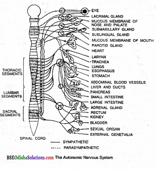
Let us take the case of a person who is angry. During anger, the action of the sympathetic system includes dilating the pupil of the eye, lifting the lid and protecting the ball, speeding up the heart rate, and raising blood pressure. There is also the cessation of digestive movements, peristaltic contractions of the stomach, and of secretion of digestive juices.
The blood that normally goes to these organs is diverted to the muscles to enable the O to face the emergency situation. All these duplicate the energy of the person. In this manner, the sympathetic system makes the O ready to face the emergency situation by helping in organizing the bodily resources to meet the situation more effectively.
Parasympathetic System:
It is a division of the autonomic Nervous System concerned with projecting and conserving the body’s resources, preserving normal functions, and maintaining a calm emotional state. It has two divisions :
- the cranial part
- The sacral part.
So it is called the craniosacral system. But here there is no such chain of parasympathetic ganglia.
The Cranial Part:
It consists of all the nerves and outlets that one, associated with the brain and the head.
The Sacral Part:
It comes from the extreme lower end of the spinal cord.
![]()
Question 7.
Describe the location of the brain.
Answer:
He conducted several experiments on rats and other animals to find out the effect of the removal of various parts of the cerebrum on psychological purposes.
- Several experiments on rats and of the problem and cerebral lessons; and
- the effects of destruction on various sizes and locations on the maze learning habits are notable.
The findings of these studies showed an intimate relationship between the difficulty of the problem and the effect of cerebral lessions. There was an increase in error while learning when cortical destructions 1 were higher. Lashley finally concluded that the quantity of the lesson was a significant factor in retardation in learning. lie found that many problems like the skill of ordinary maze learning can be acquired equally well with one part of the cortex as with another.
He further found that there was a positive relationship between the amount of cortex and the difficulty of the task that is, as the difficulty of the task increases the greater amount of cortex is required to solve it. Even when half of the cortex was removed, simple tricks were learned equally quickly, but difficult problems could not be solved.
In one experiment by Lashley (1929) adequate training was given to monkeys to open the door by handling a latch. After learning was complete the monkeys were decorticated. Some portions of the frontal lobe were removed by surgical operation, without any danger to the life of the monkey. After decortication, it was found that some portions of the brain are at least responsible for certain work.
But later he also found that money could be educated and they could relearn the previous learning after decortication. From these experimental findings, he established two principles to explain the functional localization of the brain, that is, whether the brain acts as a whole or in parts.
Principle of Equipotentiality:
Lashley was of opinion that complex learning does not depend upon the definite structure of a specific area of the cortex. On the other hand, he noted that complex learning depends upon the total organization of the cortex. The principles of equipotentiality hence mean that with some very specific exceptions.
One part of the cerebral cortex is potentially the same as another part in its functional capacity related to the learning process. In other words, the capacity of the uninjured or intact part of the brain for functioning as a substitute for other parts in case of emergency is called the principle of equipotentiality.
All parts of the cortex, therefore, are equal potential for the learning function. Thus, Lashley in his book Brain Mechanism and Intelligence (1929), has remarked, “The term equipotentiality I have used to designate the apparent capacity of the intact part of a functional area to carry out with or without reduction in efficiency, the functions which were lost by destruction as a whole.
This capacity varies from one area to another and with the character of the functions involved. It probably holds only for the association areas and for function more complex than simple sensitivity or motor coordination.”
The Principle of Mass Action:
To Lashley, the principle of mass action meant that the brain fundamentally functions as a hole. He said that the more the cortex available, the better would be the learning capacity. In this connection, he has pointed out that the animals with various amounts of their cortices removed. Showed a general reduction in sensitivity, aggressiveness, and in exploratory activity in puzzle-born learning situations in comparison to their normal counterparts.
What he intended to say is that the cerebral cortex seems to be responsible for the characteristics of one’s behavior as a whole. That is why the removal of one part of the cerebrum affects learning in a general manner. The most important generalized function of the brain is to establish associations between our various present and past experiences.
The association area of our brain contains all our experiences and it is because of this connection of experiences that our mind acts as one unit. Franz later found that the amount of brain has also got a major contribution to higher psychological activities and complex learning processes. He noticed that if a small portion of the cortex will be decorticated, it will not decrease the activities undertaken by this part.
The other parts of the brain will take charge of it and the work will be somehow managed. But if a longer part is taken away there will be a deterioration in learning and other complex mental activities. It is thus proved that more amount of cerebral cortex is needed for higher psychological processes like learning, application of intelligence and judgment, synthesizing ability, thinking, problem-solving, perception, etc.
This principle, therefore, states that the brain acts as a whole or in a mass. The principle of equipotentiality and mass action is confirmed and supported by many experimental studies on apes, monkeys, human beings, etc. These principles are also confirmed by the findings of studies on human beings whose parts of the brain were injured or damaged by disease or accident.
Analysis of these principles and the experimental findings in their support do prove that simple sensory-motor functions may show a relatively high degree of localization while complex and higher-order mental activities require less localization and more mass action. Therefore, it would probably be more appropriate to conclude that the brain acts in widespread patterns.
patterns that include many cortical areas and their connecting association fibers. Loss of any large portion of the cortex will disturb the interaction of parts and break up the usual pattern. It is the general type of function. So, we have to accept somewhere between the two. In ordinary simple work, the brain acts in part.
But in higher-order activities and higher mental processes, coordinating the integrative function of the entire brain is essential. So, in such cases the brain functions as a whole. There is evidence for the localization of sensory and muscular movements. There is some indication that the frontal lobes are concerned with the management of planned activity and that the rear half of the brain is more concerned with knowing and understanding things.
Learning seems to be an unlocalized function and adjustment of the organism for the total situation and for a goal is probably a function of the entire cerebrum. It would, therefore, be not appropriate to generalize about the principles of complete equi-potentiality of the functions of various areas of the cerebrum. Besides, the evidence derived from rats, cannot be applied with equal confidence in human organisms.
Experiments on monkeys show a much greater loss in the ability to learn when their frontal lobes were destroyed than the damage elsewhere. Some cases of human subjects also indicated that there is a considerable degree of specificity in the cortex. However, further research in this area is necessary to draw a generalized conclusion on the functional localization of the brain.
![]()
Question 8.
What are the methods applied to study the brain?
Answer:
Some of the important methods to study the localization of functional areas of the brain are discussed below:
Anatomical Method:
In this method, the anatomist attempts to find out various nerve pathways by observing the nature of neural tissues under the microscope. By this method, the anatomist not only learns the origin and termination of different nerve fibers but he also finds out what sense organs send fibers where and what parts of the brain send motor fibers to the spinal cord. This method only finds out the structure of the brain and spinal cord. The functions of the brain and the spinal cord are not traceable by this method.
Method of Extirpation or Aplation Method:
Extirpation means to destroy or cut off. In this method, a particular part of the brain is destroyed or cut off and its effect is studied on the functions of the brain or human behavior. The behavior of the organism is carefully observed and recorded. Then it cuts off a specific part of the brain and observes its effects on the behavior of the ‘O’, that is whether it changes his behavior or not.
Action Potential and Electrical Recording Method:
Neurons produce electric currents when they conduct and the potentials underlying them can be recorded with a cathode ray oscilloscope. By correlating the parts having the greatest electrical activity with the nature of the stimulus one can find out which area of the brain is involved in various types of sensations and how the body surface is represented on surfaces in the brain.
Method of Stimulation:
It is a direct method. Different parts of the brain are stimulated with mild electric currents. While making brain operations on patients an introspective report is taken from the patients on the sensations they experience while stimulation. Using this method, functions of different parts of the brain can be located.
Chemical Method:
In this method certain chemicals are used to distinguish axons from cell bodies; certain parts of neural tissues from others to observe their specific functions. Similarly, it is used to find out which axons are part of the same neurons. Otherwise, because of their complexity, it is not possible to say these things merely by looking through a microscope. Behavioral changes are also observed by using various chemicals on the external and internal parts of the brain.
Scanning Method:
Today medical science has made rapid progress, thanks to the continuous and meaningful research in the area, particularly in the West. Scanning means taking pictures intently of all parts. With the development of scamming methods, new models of X-ray machines, and computers, the functions of the brain are being studied in a very scientific and organized manner.
Through scanning, it is possible to get an X-ray picture of every millimeter of the brain. Scanning of the brain helps in knowing the damages and destruction made to the brain because of accidents. Alzheimer’s disease, Korsakoft’s Syndrome, and many other diseases like a brain tumors.
There are different types of Scanning like Cat Scans, Pet scans, and MRIs, etc.
Cat Scan:
The Cat Scan method is used when one is interested to know and analyze the minute details of the functioning of the brain of an individual. Scanning of the brain is recommended particularly when the doctor has to diagnose to patient.
Question 9.
Describe the structure of the human brain.
Answer:
The Brain:
Modem scientific psychology recognizes that is the brain, not the heart or soul which guides human behavior. Recently, technological innovations such as the electron microscope and brain scanning systems have led to an explosion in a number of new theories and tests of how the brain works. It has been found that an adult brain weighs about 1.36 kg and contains around 100 billion neurons.
The brain receives one-fifth of the blood pumped by the heart. If deprived of oxygen for 3 to 4 minutes, the brain cells are irreparably damaged. Brain investigation reveals that while some mental functions are widely distributed among different areas in the brain, man activities are highly localized.
Different areas in the brain are specialized for specific jobs. Our brain controls almost all the activities that we do, except the reflex activities which are controlled by the spinal cord.
Structure of Brain:
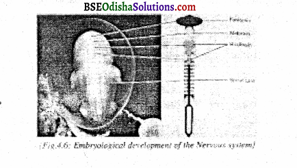
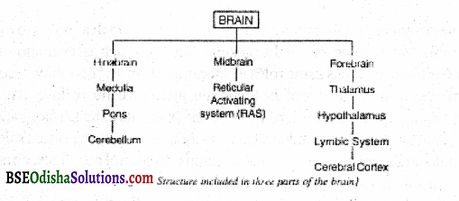
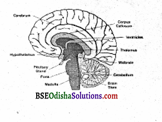
With the development of the human embryo inside the mother’s womb, the nervous system begins forming as a long, hollow tube in the back of the embryo. After three weeks of conception, cells making up the tube differentiate into a mass of neurons, most of which then develop into three major regions of the brain – the hindbrain, which is adjacent to the top part of the spinal cord, the mid-brain, which rises above the hindbrain and the forebrain, which is the uppermost region of the brain (see fig. 4.6).
Hindbrain:
It is the lowest portion of the brain located at the rear of the skull. In other words, the backside of the brain is called the hindbrain. It has three subdivisions medulla, cerebellum, and pons. The medulla begins where the spinal cord enters the skull. It is also called the medulla oblongata.
This structure is located at the lowest portion of the brain stem. It is a link between the brain stem and the spinal cord. The medulla helps to control our breathing and regulates reflexes which allow us to maintain an upright posture. It also controls some vital and autonomic functions such as respiration, circulation of blood digestion of food, etc.
It has some roles in sneezing, sleep, and coughing also. The cerebellum extends from the rear of the hindbrain, just above the medulla. It is also called the ‘Tittle brain’ because it is a miniature version of the cerebrum. Its outer surface looks grey and the interior white. It consists of two rounded structures thought to play important roles in motor coordination (Middleton & Strick, 2001).
Its vital function is to control body balance and posture. It also controls biological rhythm or perception of time. Injury to the cerebellum may lead to a lack of motor coordination, stumbling, and loss of muscle tone. When the cerebellum is damaged, movements become uncoordinated and jerky.
Extensive damage to the cerebellum even makes it impossible to stand up. It also stores the memory of movement patterns so that we do not have to concentrate on how to walk, dance, or ride a bicycle. Moreover, the cerebellum is associated with coordinating movements, controlling posture, and maintaining equilibrium.
The Pons lies between the medulla and the midbrain. It is a Latin word, which means ‘bridge’. But it does not look like a bridge. It is so named because of the bundle of nerves that passes through it. the pons region connects to the cerebellum and is involved in dreaming and waking. It contains several clusters of fibers involved in sleep and arousal (Kolb, Whishaw & Terao: 2003, 2004). Moreover, the pons transmits information about body movement and is also involved in functions related to attention, sleep, and alertness.
The Midbrain:
The shortest part of the brain is the midbrain. It is also the topmost part of the brain which is located in the central region. It is just a tube-like structure. The outside of the midbrain looks white and the inside looks grey. Through this tube, a fluid-like substance called cerebrospinal fluid passes which provides nutrition to the brain. Besides providing nutrition, it has got sensory and motor pathways. The midbrain contains primitive centers for vision and hearing and plays a key role in the regulation of visual reflexes.
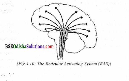
Two systems in the midbrain are of special interest – One is the reticular formation or Reticular Activating System (RAS) and the other one is the brain stem, the RAS begins in the hindbrain and ascends through the region of the midbrain into the lower part of the forebrain. It is a network of neurons crossing each other.
The size of RAS is just like a small finger of a man. RAS has two parts ascending reticular system and descending reticular system. The ascending reticular system sends impulses to the cerebral cortex and the descending system sends impulses downward to the RAS. RAS also receives impulses from the cerebral cortex.
Electrical stimulation of RAS awakens sleeping animals. If the RAS is damaged, the animal may not die but will sleep forever. This kind of sleep is called comatose or simply coma. Once it is destroyed, it does not recover. RAS also acts as a relay station for emotional behavior. The RAS is less activated during sleep. It is connected with the cerebrum by receptors and effectors. Further, it plays an important role in selective attention and filtering of information through learning.
The brain stem is so-called because it looks like a stem (Carlson, 2001). It is the most ancient part of the brain. The brain stem connects with the spinal cord at its lower end and then extends upward to encase the reticular formation in the midbrain. Clumps of cells in the brain stem determine alertness and regulate basic survival functions, such as breathing, heartbeat, and blood pressure.
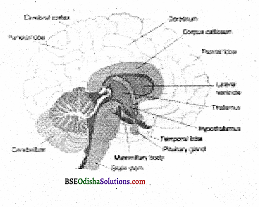
The Forebrain:
It is the most important part of the brain. Virtually it possesses all the parts concerned with the perception, and coordination of behavior patterns including those of emotion, motivation, learning, memory, language, and thinking. The significant parts of the forebrain are the thalamus, hypothalamus, limbic system, and cerebrum.
Thalamus:
Thalamus is located almost in the exact center of the human brain. It consists of an egg-shaped cluster of neurons. Thalamus connects the cerebrum with peripheral parts of the body. All the nerves come and go through it. So it is an important relay station for incoming sensory impressions from all parts of the body. Further, it is called the central switchboard of the brain.
The main function of the thalamus is to send incoming sensory impulses to respective parts of the cerebral cortex. Most neural input to the cerebral cortex goes through the thalamus. Thalamus is also involved in controlling sleep and attention in coordination with other brain structures, including RAS. When the cortex wants to inhibit or control certain automatic activities, it sends the impulses to the thalamus.
Basal Ganglia:
Just above the thalamus and under the cerebral cortex lie large clusters of neurons called basal ganglia. The basal ganglia work with the cerebellum and the cerebral cortex to control and coordinate voluntary movements. These large clusters of neurons or ganglia enable people to engage in habitual behaviors such as riding a bicycle. People with damage to basal ganglia suffer from either unwanted movement, such as constant writing or jerking of limbs, or too little movement, such as the slow and deliberate movements of those with Parkinson’s disease.
Hypothalamus:
Hypothalamus is a small forebrain structure located just below the thalamus. It lies at the base of the cerebrum. It regulates the functioning of the Autonomic Nervous System. Hypothalamus monitors three pleasurable activities eating, drinking, and sex – as well as emotion, stress, and reward. It also directs the endocrine system. Hypothalamus acts as a regulator of the body’s internal state.
It also plays an important role as an integrative location for handling stress (Hayashi and others, 2004). Much of the integration is accomplished through the hypothalamus’s action on the pituitary gland, an important endocrine gland located just below it. If certain areas of the hypothalamus are stimulated electrically, a feeling of pleasure results.
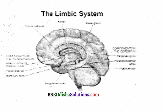
The Limbic System:
The limbic system is composed of a group of structures that is found in all mammals, sometimes called the old brain. Only mammals and reptiles have limbic systems. It is a ring-like structure having several other structures. The limbic system is structurally interconnected with the hypothalamus. So it is involved in the drives of hunger, sex, aggression, and some of the behaviors regulated by the hypothalamus.
The three principal structures in the limbic system are the amygdala, hippocampus, and septum. The amygdala has an important role in aggression. It is involved in memory, emotions, and certain basic motivations. The damage to the amygdala causes an animal to be less fearful, over-curious, hypersexual, and more exploratory.
![]()
Question 10.
What is the endocrine gland and discuss the functions of the endocrine system?
Answer:
The endocrine system is a set of glands that regulate the activities of certain organs by releasing their chemical products into the bloodstream, like the nervous system, it plays a crucial role in our behavior and development previously, the endocrine system was considered separate from the nervous system.
However, today neuroscientists know that two systems are interconnected. The endocrine system consists of ductless glands which secret complex chemical substances called hormones, directly into the bloodstream. The human body is not only under the control of the nervous system but also of a complementary system of hormones.
This system is controlled by Autonomic Nervous System (ANS) activity and like ANS, it is not under conscious control but regulated by the body itself. It helps in maintaining bodily homeostasis. Hormones are involved in different bodily functions and behaviors. They influence body growth, sexual development arousal, mood, and metabolism.
Endocrine or ductless glands are stimulated in 3 ways:
- by chemical level in the bloodstream,
- other hormones and
- nerve impulses from the Bram
Once secreted into the blood, hormones are promoted it’s their bodily targets. This system not only sustains our slow and continuous bodily processes but also helps us to respond to crises. During an emergency, the Iannone ‘adrenaline’ is released into the bloodstream, energizing our body for quick defensive action for ‘ fight’ or ‘ flight’.
‘Hormones’ are generally called ‘the messengers of life’ because their influence is diverse but specific (Carpo 1988). Different hormone factories ‘sites of our body produce chemicals that influence a variety of bodily processes. A small structure of the limbic system, the hypothalamus, is the brain center in charge of the endocrine system.
In the hypothalamus, specialized cells receive messages from other brain cells committing to release a number of different chemicals. These chemicals influence the adjacent pituitary gland, the so-called master gland which can either stimulate or inherit the release of hormones from other glands.
The functions of the Endocrine System:
The important functions of the endocrine system are discussed below:
- The pituitary gland
- Thyroid gland
- Parathyroid
- Adrenal gland
- The pancreas
- The Gonads
The Pituitary Gland:
Although the pituitary gland is situated within the cranium, still it is a part of the endocrine system, rather than the nervous system. This gland is popularly known as the master gland. It is located in a small bony hollow at the base of the brain. Centre of the skull. The size of the pituitary gland is very small. But control growth and regulates other glands. This pea-sized gland is controlled by the hypothalamus.
The pituitary gland has:
- anterior
- posterior pituitary secretions.
Anterior:
The anterior pituitary secretions help growth. Hypersecretion of it causes gigantism and a rugged personality.
Posterior pituitary secretions:
The posterior pituitary gland hormone raises blood pressure, regulates metabolism, and increases the contraction of smooth muscles in the intestine and uterus.
Thyroid Gland:
The thyroids are located in the neck at either side of the ‘Adam’s apple’. These glands produce thyroxin, which influences the body’s metabolism rate. It also helps to control the rate of physical growth and influences the structure and functions of the nervous system.
![]()
Question 11.
What is the cerebrum and describe how does the brain function?
Answer:
The Cerebrum:
The cerebrum or cerebral cortex is more highly developed in humans than in any other animals. The cerebral cortex is divided into two halves or hemispheres. It is the highest region of the forebrain and is the most recently developed part of the brain in the evolutionary scheme. In humans, the cerebral cortex covers the lower portions of the brain like a large cap.
The cortex is greatly convoluted with lots of grooves and bulges which considerably enlarge its surface area. It is connected with other parts of the brain. Literally, millions of axons connect the neurons of the cerebral cortex with those located elsewhere in the brain.
The cortex looks gray since it consists largely of cell bodies and unmyelinated fiber. A very depressed or fissure divides the cerebrum into two equal halves – the left hemisphere and the right hemisphere. The left hemisphere is connected with the right-hand side of the body. The right hemisphere is connected with the left-hand side of the body.
The two hemispheres are connected with each other by a thick fiber bundle known as the corpus callosum. Each hemisphere is divided into four parts or lobes by two fissures – the frontal lobe, the parietal lobe, the temporal lobe, and the occipital lobe. However, the occipital lobe is not clearly demarcated. It is located at the rear of the brain.
The frontal lobe is that portion of the cortex behind the forehead which is involved in the control of voluntary muscles, intelligence, and personality. The frontal lobes of humans are especially large when compared with those of other animals. This lobe is primarily responsible for the planning, execution, and control of movements.
Without intact frontal lobes, humans are emotionally shallow, -5 distractible, listless, and insensitive to social contexts (Hopper & Teresi, 1992). Individuals with frontal lobe damage become so distracted by irrelevant stimuli that they often cannot carry out some basic directions. An important part of the frontal lobe is the prefrontal cortex, which is at the front of the motor cortex.
It is believed to be involved in higher cognitive functions such as planning and reasoning (Manes & Others, 2002). Neuroscientists refer to the prefrontal cortex as an executive control system because of its role in monitoring and organizing thinking (Owen, 1997). The parietal lobe is located at the top and towards the rear of each hemisphere.
It is involved in registering spatial location, attention, and motor control. This cortex receives information from the somatic senses. Therefore, this area is specialized for touch, pressure, and pain. On the whole, it controls incoming sensory information. The portion of the cerebral cortex just above the ears is the temporal lobe. It is involved in hearing, language processing, and memory.
The temporal lobes have a number of connections to the limbic system. Individuals with damage to the temporal lobes can not file experiences into long-term memory. The area of recognition of music is situated in this area. If this area is destroyed, an individual may lose the taste sensation.
The occipital lobe is located at the back portion of the brain. Its shape is triangular. It is the seat of the visual sensation. The most important functional area located in this lobe is the visual area. The retina is connected to this area. A stroke or wound in the occipital lobe can cause blindness or wipe, out a portion of the person’s visual field.
In the cerebral cortex, there are some areas that are not directly concerned with sensory or motor functions. These are called association areas. Each lobe is having an association area. They play significant roles in various sensory systems and in transmitting sensory input to programmers for motor output. Further, the association areas are involved in complex cognitive activities such as thinking, reasoning, learning, remembering, etc.
How does the brain function?
Very often, the question arises whether or not the brain functions as a ‘whole’. Physiological psychologists are trying to answer this question since the days of Johannes Muller. There are differences in views. A group of experts believed that each part of the cerebrum had a definite function. Another group believed that parts of the brain are functionally interchangeable. Modem findings indicated that the brain functions in parts as well as as a whole.
The experimental findings of Franz and Lashley on localization are expressed in two theories:
- the theory of equipotentiality and
- the theory of mass action.
Theory of Equipotentiality:
The theory of equipotentiality suggests that all parts of the cortex are equal potential enough for simple learning functions. Lashley has conducted a good number of studies on animals. According to him, complex learning does not depend upon the definite structure of a specific area of the cortex, rather, complex learning depends upon the total organization of the cortex.
This principle suggests that with some very specific exceptions, one part of the cerebral cortex is potentially the same as another part in its functional capacity related to the learning process. Therefore, all parts of the cortex are equal potential for learning function.
The Principle of Mass Action:
The principle of Mass Action reveals that the brain fundamentally functions as a whole. The more parts of the cortex an available, the better would be the learning capacity. In his study, Lashley found that animals having decorticated cortex demonstrated a general reduction in sensitivity, exploratory activities, and aggressiveness. The removal of any part of the brain affects the learning process.
Later, Franz found that the amount of brain has got major contributions to higher psychological activities and complex learning processes. The principles of Mass Action and Equipotentiality were confirmed and supported by many experimental studies on apes, monkeys, human beings, etc.
These principles were also confirmed by the findings of the studies on human beings whose parts of the brain were injured or damaged by disease or accident. Probably it would be more appropriate to conclude that the brain acts in widespread patterns, which include many cortical areas and their connecting association fibers.
Damage to the large portion of the cortex would certainly disturb the interaction of parts and break up the usual pattern. It is true that the brain acts in part in ordinary simple work. But in higher mental activities and higher mental processes, the coordinating and integrative function of the entire brain is necessary. In such cases, the brain functions as a whole. However, to draw a generalized conclusion on the functional localization of the brain, further research in this area is essential.
![]()
Question 12.
What is the peripheral Nervous System?
Answer:
The peripheral nervous system is composed of all the neurons forming the nerve fibers that connect the CNS to the rest of the body. It consists of sensory and motor neurons that transmit messages to and from the central nervous system. Our brain will be isolated from the world without a peripheral nervous system. This system has two divisions.
- Somatic Nervous System.
- Autonomic Nervous System.
These are described below:

Somatic nervous system:
The somatic nervous system is the first part of the peripheral nervous system. It is under voluntary control and regulates the actions of the skeletal muscles of the body. The somatic nervous system has both sensory neurons and motor neurons. The sensory neurons mn from our sense organs toward the central nervous system for perception and learning etc.
On the other hand, the motor neurons carry messages from the brain to the striped muscles of the body for activities. Action like moving our legs or hands, running, jumping, and riding is possible by the somatic nervous system. On the whole, the somatic nervous system controls the striped muscles of our body.
Autonomic Nervous System (ANS)
The very word autonomic means self-regulating or independent. ANS is the second part of the peripheral nervous system. It is involuntary and governs activity, which is not normally under the direct control of the individual. The ANS works even when we are asleep. ANS sustains basic life processes.
The ANS operates constantly, regulating bodily processes we do not usually control consciously, such as respiration, digestion, and arousal. On the whole, the autonomic nervous system is called as autonomic because many of the activities it controls are autonomous or self-regulating and continue even when a person is asleep or unconscious.
The autonomic nervous system (ANS) is subdivided into the sympathetic and parasympathetic nervous systems. Both these divisions have survival functions for which the autonomic nervous system is called as survival nervous system. The ANS is intimately connected with the spinal cord.
The sympathetic division tends to act as a unit. It is exciting in emotional situations. The sympathetic division governs responses to stress in emergencies when action must be quick and powerfully, energized. This is the ‘fight or flight response system. In short, the sympathetic nervous system is the division for emergency survival.
For example, during emotional excitement, digestion is stopped, heart rate is increased, blood flowing to the internal organs is directed to the skeletal muscles and the endocrine system is stimulated to release several chemicals which increase the effectiveness of the entire motor system. The sympathetic division energizes us to respond to a stressor quickly.
But the parasympathetic division monitors the routine operation of the body’s internal functions. It is concerned with projecting and conserving the body’s resources, preserving normal functions, and maintaining a calm emotional state. This division returns the body to calmer functioning after sympathetic arousal. Unlike the sympathetic division, the parasympathetic division tends to affect one organ at a time.
In short, whereas the sympathetic system is activated during violent emotions, the parasympathetic system is dominant and active during normal times or during quiescence. Activation of this system slows the heartbeat, lowers blood pressure, and conserves as well as protects bodily resources. The sympathetic and parasympathetic systems do not compete with one another, rather they function in a coordinated manner.
Points to remember
Question 1.
What is Neuron? Describe the structures and functions of a Neuron, How does it differ from a cell?
Building Blocks of the Nervous System:
Neurons are basic units of the nervous system. On the whole, the way the neurons function reflect the major characteristics of the nervous system.
Basic Structure of a Neuron:
In general, every neuron has the following structures.
- Cell body or soma
- Dendrites
- Axon and
- Terminal buttons or axon terminals
![]()
Question 2.
Central Nervous System and Structure and Function.
Answer:
The central nervous system is well protected in the bony case of the skull and spinal column. It is divided into the spinal cord and the brain.
Question 3.
State the structure and function of the autonomic nervous system.
Answer:
Autonomic Nervous System:
The anatomical distinction between the central nervous system and the autonomic nervous system lies in that the nerve fibers of the A.N.S. have always a junction with another neuron outside the brain or spinal cord on the way to muscles or glands.